


What Is Apical Resection? Cost and Post-Operative Care
Don't put off your smile. Message us now to discuss your treatment options with our expert dentists.
WhatsAppIn clinical dentistry, the foundational principle is always to preserve the patient’s natural teeth and avoid extraction whenever biologically feasible. To achieve this, root canal therapy (endodontic treatment) is routinely implemented when the internal tooth pulp becomes necrotic or infected. However, due to highly calcified root anatomies, inaccessible accessory canals, or extremely resistant bacterial strains, traditional root canal treatments can sometimes fail. This failure results in chronic infections, granulomas, and bone-resorbing cysts forming at the tooth root tip (apex).
While extraction historically seemed like the only viable solution for teeth reaching this stage, modern oral surgery offers a highly successful last line of defense to salvage natural teeth: apical resection (root-end surgery / apicoectomy). This microsurgical procedure accesses the infected root tip directly by creating a tiny window through the overlying jawbone, allowing the surgeon to excise the root-end, curette the cyst, and secure the tooth’s functional presence in the arch. This comprehensive YMYL master guide explores what is apical resection, how the surgery is performed, 2026 apical resection cost factors, and vital guidelines for managing your after apical resection recovery.
Table of Content
- What is Apical Resection?
- How is Apical Resection Done?
- What to Expect After Apical Resection: Swelling and Healing Timelines
- Critical Things to Consider After Apical Resection
- Apical Resection Success Rate and Infection Recurrence Risks
- Performance Matrix: Apicoectomy vs. Extraction and Implant
- How is the Investment Determined: Apical Resection Cost
- Frequently Asked Questions
What is Apical Resection?
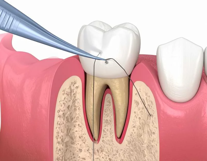
In professional endodontic surgery, the precise scientific answer to what is apical resection is this: It is the surgical excision of a tooth's root tip (apex) along with the surrounding chronically infected granulomatous or cystic tissues, performed under local anesthesia by creating a micro-window through the alveolar bone.
Once the compromised root-end is cut away, a biocompatible material (typically MTA - Mineral Trioxide Aggregate) is packed retrogradely (retrograde filling) into the canal entry to seal the root airtight. This procedure’s primary intent is to eliminate persistent subgingival bacterial reservoirs that cannot be reached via standard root canal approaches, bypassing tooth extraction entirely.
How is Apical Resection Done?
Conducted precisely inside our clinical surgical suites through a coordinated approach between our oral surgeons and endodontists, the sequence of how is apical resection done proceeds through these painless phases:
- 3D Volumetric Imaging (CBCT): Before making any incisions, a 3D cone-beam computed tomography scan is captured to map the precise volume of the bone lesion and locate adjacent anatomical landmarks, such as the mental nerve in the lower jaw or the maxillary sinus cavities in the upper jaw.
- Profound Local Anesthesia: To guarantee a completely painless surgical experience, local anesthetic agents are administered to fully switch off sensory pathways around the target tooth and bone quadrant.
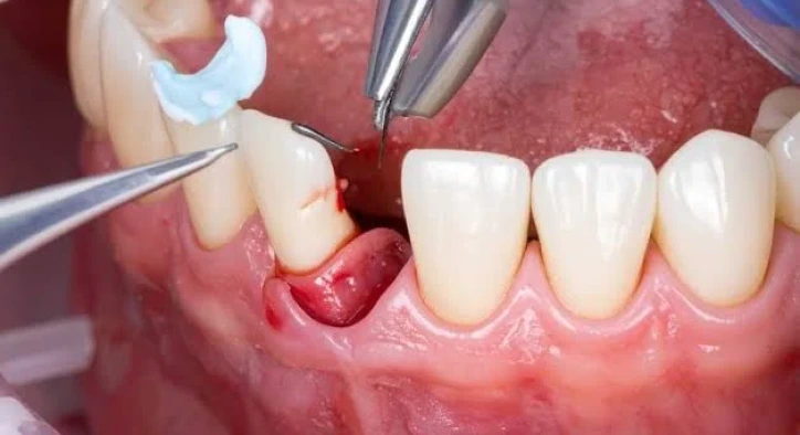
- Accessing the Root Apex: A small, aesthetic flap is incised in the gingival tissue and gently reflected to expose the underlying alveolar bone. Using high-speed microsurgical handpieces under continuous saline irrigation, a tiny micro-window is milled through the bone to expose the diseased root tip.
- Excision of the Root-End & Cyst Curettage: The apical 3 mm of the root tip (where microscopic accessory canals housing bacteria are most concentrated) is carefully sectioned and removed. All surrounding inflammatory tissues, granulomas, or cysts are meticulously curetted and cleared from the bone cavity.
- Retrograde Filling & Closure: A retrograde cavity preparation is milled into the sectioned root face and packed with a biocompatible MTA seal. If the bone defect left by the cyst is extensive, high-grade bone powder (bone graft) is placed to support rapid bone regeneration. Finally, the gum flap is repositioned and secured with micro-sutures.
What to Expect After Apical Resection: Swelling and Healing Timelines
The post-operative healing phase is critical to secure the long-term biological attachment of the root structure.
Apical Resection Recovery Time
Initial superficial healing of the soft tissue flap and the closing of the gum margin is completed in 7 to 10 days, at which point the micro-sutures are cleared. However, the internal regeneration process required for the empty bone cavity to fill completely with new, healthy trabecular bone takes an average of 6 to 9 months. This internal bone remodeling proceeds quietly without any noticeable physical sensation for the patient.
Swelling After Apical Resection
Experiencing mild to moderate swelling after apical resection on the treated cheek or lip quadrant is a completely normal, physiological response to surgical manipulation. This postoperative edema peaks within the first 48 hours and begins to resolve rapidly starting on day 3. Applying an external cold compress over the cheek for 10 minutes on and 10 minutes off during the first 24 hours is highly effective at minimizing swelling.
Critical Things to Consider After Apical Resection
To protect the healing soft tissue flap and prevent bacterial contamination, adhere to these vital things to consider after apical resection rules:
- Zero Muscle Tension: Avoid pulling your lips or cheeks outward to inspect the stitches in a mirror. Any stretching of the facial muscles creates immediate tension on the micro-sutures, which can tear the flap borders open.
- Dietary Adjustments: For the first 48 hours, avoid extremely hot, cold, spicy, or crunchy foods. Maintain a lukewarm, soft diet (such as purees, yogurts, and lukewarm broths) and chew exclusively on the opposite side of your mouth.
- Aqueous Hygiene & Brushing: Do not brush the surgical area on the day of the procedure. From day two, all other teeth must be brushed and flossed normally. Gently clean the treated gumline using an ultra-soft post-operative toothbrush, and rinse gently with the prescribed chlorhexidine mouthwash without aggressive swishing.
- Limit Physical Strain: Avoid intense workouts, heavy lifting, sauna visits, or hot baths for the first 3 days post-surgery, as elevated systemic blood pressure can trigger secondary bleeding.
Apical Resection Success Rate and Infection Recurrence Risks
When cases are carefully selected and executed using micro-surgical techniques, endodontic surgery boasts a highly predictable prognosis.
- Apical Resection Success Rate: Modern clinical tracking shows that apicoectomies completed with biocompatible retrogradely placed seals achieve a success rate of 85% to 92%. The primary factor driving long-term survival is securing a complete, airtight seal at the root-end.
- Infection Recurrence After Apical Resection: Though rare, infection recurrence after apical resection can occur. Primary causes of failure include incomplete apical seals, unrecognized micro-cracks along the vertical axis of the root, or secondary periodontal-endodontic communication. If recurrence occurs and progressive bone loss is confirmed, the final clinical step is tooth extraction followed by a dental implant.
Performance Matrix: Apicoectomy vs. Extraction and Implant
Compare the biological and structural pathways of both clinical choices below:
|
Technical & Clinical Criteria |
Apical Resection Procedure |
Tooth Extraction & Dental Implant |
|
Natural Tooth Preservation |
Yes (Your natural tooth remains functional) |
No (The natural tooth is completely lost) |
|
Surgical Scope |
Microsurgical (Focused solely on the root tip) |
Full extraction followed by bone-embedded post |
|
Average Appointment Time |
30 to 45 Minutes |
Extraction (15 min) + Implant placement (20 min) |
|
Soft Tissue Healing Window |
7 to 10 Days (Concludes with suture removal) |
Requires 3 months for bone integration |
|
Aesthetic and Functional Transition |
Immediate (The dental crown remains unaltered) |
Temporary prosthetic phase until final crown placement |
|
Routine Bone Grafting Need |
Indicated only for very large bone cavities |
Highly common to preserve extraction socket volume |
How is the Investment Determined: Apical Resection Cost
Stating a flat fee for "apical resection cost" prior to a physical assessment is clinically inaccurate. Final treatments track the baseline indices of the Turkish Dental Association alongside these specific variables:
- The Anatomical Position of the Tooth: Apicoectomies on single-rooted front teeth are structurally less complex compared to multi-rooted posterior molars, directly influencing the surgical fee structure.
- Lesion Scale and Biomaterial Requirements: If the cyst has created a large bone cavity, the volume of high-grade bone graft (bone powder) and protective collagen membranes required to stabilize the site will adjust the final budget.
- The Choice of Retrograde Sealing Materials: Utilizing premium-tier biocompatible materials such as MTA to secure a permanent, hermetic root-end seal influences material costs.
Frequently Asked Questions
Will I feel any pain during the apical resection surgery?
No, absolutely not. The entire procedure is conducted under profound local anesthetic blocks that fully switch off local pain pathways, ensuring that you feel no discomfort during the surgery. You will only feel slight tactile pressure or water spray from the micro-instruments; any sharp pain is biologically blocked.
Is there intense pain after the anesthesia wears off?
Once the local anesthesia subsides, it is normal to experience a mild, dull post-operative ache or tightness around the surgical site. This discomfort is highly manageable and is easily controlled with the anti-inflammatory painkillers prescribed by your surgeon. Most patients comfortably return to their normal routines the very next day.
How long does a tooth last after undergoing an apical resection?
When executed with high precision and backed by solid home care, a tooth treated with apical resection can function comfortably in your mouth for a lifetime. The ultimate longevity of the tooth depends on maintaining excellent daily oral hygiene to protect the surrounding gums from periodontal disease.
When are the sutures removed after an apical resection?
If non-resorbable micro-sutures were used, they are gently and painlessly removed by your surgeon exactly 7 to 10 days after the procedure, once the soft tissue flap borders have stabilized.
About the Author