




What Is Oral Cancer? Symptoms, Stages, and Current Treatment
Don't put off your smile. Message us now to discuss your treatment options with our expert dentists.
WhatsAppThe oral cavity serves as the primary operational gateway for vital human physiological functions, including speech, mechanical chewing, deglutition (swallowing), and respiration. Comprising the lips, tongue mobile surfaces, buccal mucosa (inner cheeks), gingiva, hard and soft palates, and the floor of the mouth, this delicate ecosystem is continuously exposed to external microbes, thermal fluctuations, and chemical inputs. Under normal conditions, minor intraoral abrasions, ulcers, or aphthous lesions heal spontaneously within a few days through homeostatic immune actions. However, occasionally, chronic cellular irritation or intense exposure to carcinogens can cause genetic mutations in the mucosal epithelium, inducing uncontrolled cell division.
Classified as a major subset of head and neck oncology, these malignant structural growths are termed oral cancer (cancer of the oral cavity). Predominantly manifesting as oral squamous cell carcinoma (OSCC), this pathology is highly treatable when intercepted during early cell colonization, but it introduces severe life-threatening prognosis if left to advance systematically. This comprehensive YMYL master guide explores what is oral cancer, unmasks internal oral cancer causes, provides tracking for vital oral cancer symptoms, and deciphers modern multidisciplinary oral cancer treatment algorithms.
Table of Content
- What is Oral Cancer?
- Oral Cancer Causes: Identifying the Structural Triggers
- Oral Cancer Symptoms: Recognizing the Warning Signs
- Oral Cancer Stages and Diagnostic Classification
- Prognosis Truths: Can Oral Cancer Kill You?
- Contemporary Protocols: Modern Oral Cancer Treatment
- Diagnostic Matrix: Simple Mouth Ulcer vs. Oral Cancer
- Frequently Asked Questions
What is Oral Cancer?
In contemporary clinical oncology, the precise scientific answer to what is oral cancer is this: It is the malignant transformation and uncontrolled clonal proliferation of squamous epithelial cells that line the mucosal surfaces of the oral cavity.
Oral malignancies are highly invasive. If left unchecked, the lesion will cross tissue boundaries, migrating deep into underlying muscle matrices, eroding adjacent jawbone structures, traveling via lymph nodes to the cervical neck chains, and eventually metastasizing to distant organs like the lungs. Routine diagnostic dental screening is therefore crucial not just for discovering dental decay, but for identifying these asymptomatic, early-stage lesions.
Oral Cancer Causes: Identifying the Structural Triggers
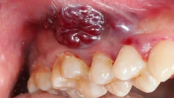
While a single genetic factor is rarely isolated, clinical data confirms that certain carcinogens and continuous physical friction break down cellular repair mechanics, acting as primary oral cancer causes:
- Chronic Tobacco Exposure: Cigarettes, cigars, pipes, and smokeless chewing tobacco are the leading causes of oral cavity malignancies. The complex chemical carcinogens in tobacco cause direct DNA adduct mutations within the epithelial lining.
- Alcohol Synergism: Chronic, heavy alcohol consumption dissolves protective mucosal lipids, accelerating the absorption of localized carcinogens into the basal cell layer. Combining alcohol and tobacco creates a synergistic effect, multiplying oral cancer risk approximately fifteenfold.
- Human Papillomavirus (HPV) Infection: High-risk viral strains (specifically HPV-16 and HPV-18) are strongly linked to the rising incidence of malignancies located at the base of the tongue, tonsils, and posterior oropharyngeal borders.
- Chronic Mechanical Friction: Defective, ill-fitting dentures with rough borders, sharp fractured teeth, or broken restorations create continuous physical trauma against the tongue or inner cheeks for years. This persistent injury forces rapid cellular turnover, which can spark malignant transformation.
- Actinic Radiation Exposure: Excessive ultraviolet (UV) solar radiation is the primary environmental cause of lower lip cancers, typically developing from a precursor lesion known as actinic cheilitis.
Oral Cancer Symptoms: Recognizing the Warning Signs
Differentiating between a simple, self-limiting mouth ulcer and an early-stage malignancy is critical for patient survival. The most prominent clinical oral cancer symptoms include:
- Persistent Ulceration: Any intraoral sore, fissure, or raw erosion that fails to achieve complete structural healing within 14 days (2 weeks) despite conservative treatments is a primary warning sign of oral cancer.
- Leukoplakia and Erythroplakia: The presence of persistent white patches (leukoplakia) that cannot be scraped off, or velvety, bright red areas (erythroplakia) across the mucosal lining signify high-risk precancerous changes that require immediate biopsy.
- Indurated Masses and Spontaneous Bleeding: The development of a firm, hard lump or thickness beneath the soft tissue matrix, or unexplained, spontaneous bleeding from localized gingival tissues.
- Functional Mobility Limitations: Loss of normal tongue flexibility, difficulty chewing or moving the jaw (trismus), and a persistent sensation of a lump in the throat during swallowing.
- Voice Changes and Otalgia: Unexplained persistent hoarseness or vocal tone shifts, combined with chronic, unilateral ear pain (referred otalgia) triggered by shared cranial nerve pathways.
Oral Cancer Stages and Diagnostic Classification
Treatment path planning relies on staging using the TNM (Tumor size, Node involvement, Distant Metastasis) framework. The clinical breakdown of oral cancer stages is categorized as follows:
- Stage 0 (Carcinoma in Situ): Malignant cells are confined strictly within the superficial epithelial layer, showing zero invasion into deeper tissues. Curability approaches 100%.
- Stage I: The primary tumor measures 2 cm or less in its greatest dimension and shows no spread to local lymph nodes or distant sites (Early Stage).
- Stage II: The tumor grows larger, measuring between 2 cm and 4 cm, but remains localized with zero lymph node involvement.
- Stage III: The primary tumor exceeds 4 cm, or is any size but has metastasized to a single ipsilateral cervical lymph node measuring 3 cm or less.
- Advanced Oral Cancer (Stage IV): Subdivided into categories A, B, and C. In advanced oral cancer, the primary tumor invades adjacent cortical jawbone structures, deep facial muscles, or external skin profiles, or spreads to multiple extensive regional lymph nodes. Stage IVC represents distant metastasis, where tumor clusters travel through the bloodstream to establish secondary sites in organs like the lungs, liver, or bones.
Prognosis Truths: Can Oral Cancer Kill You?
During the initial diagnostic consultation, the most distressing question patients ask is can oral cancer kill you?
The medical reality is straightforward: Survival depends entirely on the stage at diagnosis. Oral cancer is an aggressive disease that, if left untreated, will invade deep vital structures and cause systemic failure, making it potentially fatal.
However, when diagnosed early (Stages I and II), the 5-year survival and complete cure rate is exceptionally high, ranging from 80% to 90%. Conversely, if the pathology reaches lymph node involvement or presents as advanced oral cancer (Stage IV), the 5-year survival rate falls to roughly 30% to 40%. This drastic statistical drop underscores why any suspicious lesion lasting over 2 weeks requires an immediate diagnostic biopsy.
Contemporary Protocols: Modern Oral Cancer Treatment
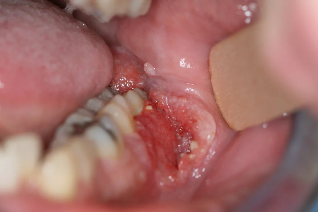
Managed by a multidisciplinary head and neck oncology board containing maxillofacial surgeons, oncologists, and microvascular reconstructive specialists, modern oral cancer treatment protocols include:
1. Radical Surgical Resection
Surgical excision remains the primary cure for oral cavity tumors. The surgeon resects the tumor mass alongside a wide margin of healthy tissue to ensure clear borders. If the tumor invades the jaw, a partial jawbone resection (mandibulectomy/maxilectomy) is performed. Concurrently, a selective or radical neck dissection is executed to clear the regional lymph node chains. Reconstructive surgeons immediately repair the defect using microvascular free flaps harvested from the patient's fibula or forearm, restoring functional speech and swallowing mechanics.
2. Adjuvant Radiotherapy and Chemotherapy
For advanced or high-risk cases (Stages III and IV), focused high-energy radiation beams (radiotherapy) are directed at the surgical site to eradicate any remaining microscopic cancer cells. Systemic drugs that arrest cell division (chemotherapy) are often delivered concurrently with radiation to maximize the treatment's therapeutic effect.
Diagnostic Matrix: Simple Mouth Ulcer vs. Oral Cancer
Review this clinical matrix to help differentiate between a benign aphthous ulcer and a suspicious malignant growth:
|
Clinical Criteria |
Benign Aphthous Ulcer (Canker Sore) |
Suspected Oral Cancer Lesion |
|
Healing Duration |
Resolves spontaneously within 7–10 days. |
Persists beyond 14 days, showing zero healing progress. |
|
Tissue Texture & Borders |
Soft to the touch, centered with white/yellow slough. |
Indurated (firm/hard) with irregular, raised borders. |
|
Pain Characteristics |
Sharp, intense pain during the initial days. |
Completely painless at onset, progressing silently. |
|
Hemorrhage Potential |
Rarely bleeds unless directly traumatized. |
Exhibits frequent, spontaneous bleeding. |
|
Surrounding Mucosa |
Appears normal, localized halo inflammation. |
Framed by persistent white (leukoplakia) or red patches. |
|
Cervical Lymph Nodes |
Remain unaffected or present with soft, tender swelling. |
Accompanied by hard, fixed, painless neck lumps. |
Frequently Asked Questions
What screening actions can I take at home for early oral cancer detection?
You should perform a self-screening monthly in front of a mirror under bright lighting. Lift your lips to check the inner mucosal linings, retract your cheeks, and inspect the entire surface of your tongue—including the lateral borders and underneath. If you discover white or red patches, hard tissue lumps, or raw sores that have persisted for more than two weeks, consult an oral and maxillofacial surgeon immediately.
Can individuals who have never smoked develop oral cancer?
Yes, they can. While chronic tobacco exposure remains the primary statistical risk factor, roughly 25% of all diagnosed oral cancer patients have no history of tobacco or heavy alcohol use. In these cases, the disease is frequently driven by high-risk oncogenic HPV strains, nutritional deficiencies, or prolonged mechanical friction from sharp, broken teeth or defective denture borders.
Does performing a tissue biopsy cause the cancer to spread?
No. This is a dangerous medical myth with no scientific basis. Taking a small tissue sample (biopsy) under local anesthesia does not cause a tumor to spread or worsen. A biopsy is the only definitive scientific method available to confirm a diagnosis, identify the specific cell type, and map an accurate cancer treatment plan. Delaying a biopsy out of fear allows the primary tumor to grow unchecked and migrate into the jawbone and lymph systems.
Can routine dental care be performed after oral cancer treatments?
If your oral cancer treatment included radiation therapy (radiotherapy) to the jaw quadrants, the local capillary network becomes permanently altered, significantly reducing the bone's capacity to heal. Performing routine extractions or placing dental implants in a radiation-exposed jaw years later can trigger a severe condition called osteoradionecrosis (radiation-induced bone death). Consequently, all post-radiation dental care must be managed using strict surgical protocols under the direct guidance of a maxillofacial surgeon and your radiation oncologist.
About the Author