


What Is a Jawbone Cyst? Symptoms, Surgery, and Treatment
Don't put off your smile. Message us now to discuss your treatment options with our expert dentists.
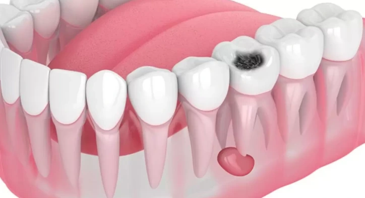
WhatsAppHuman jaw architecture comprising the upper jaw (maxilla) and lower jaw (mandible) is a complex skeletal structure housing tooth roots, neurovascular bundles, and sinus cavities. However, due to tissue remnants around impacted wisdom teeth, untreated deep dental decay reaching the root apex, or embryonic developmental anomalies, fluid-filled pathological sacs can form inside the jawbone.
In oral and maxillofacial surgery, these intraosseous sacs are termed a jawbone cyst. Expanding silently within the bone over years without early pain, these pathologic cavities cause localized bone resorption, thinning the cortical bony walls. Often discovered incidentally during routine panoramic radiographs or 3D cone-beam computed tomography (CBCT) scans, jaw cysts can be completely cured through proper surgical protocols. This comprehensive YMYL master guide explores what is a jawbone cyst, analyzes what causes a jawbone cyst, details key jawbone cyst symptoms, explains lower jawbone cyst surgery.
Table of Content
- What is a Jawbone Cyst? Biological Structure
- What Causes a Jawbone Cyst? Classifications and Triggers
- Jawbone Cyst Symptoms: Identifying Clinical Signs
- Is a Jaw Cyst Dangerous?
- How to Shrink a Jaw Cyst?
- Surgical Removal: Lower Jawbone Cyst Surgery
- Post-Operative Recovery: Jaw Cyst Surgery Recovery
- Comparative Matrix: Enucleation vs. Marsupialization
- Frequently Asked Questions
What is a Jawbone Cyst? Biological Structure
In oral surgery literature, the exact scientific answer to what is a jawbone cyst is this: It is a fluid- or semi-fluid-filled pathologic cavity lined by epithelium, developing inside the maxilla or mandible, which progressively expands by resorbing the surrounding alveolar bone.
As a jaw cyst expands mechanically, it exerts hydrostatic pressure on surrounding bone cells (osteoclasts). This pressure stimulates localized bone resorption. If left untreated, a large cyst can thin the jawbone to the fragility of an eggshell.
What Causes a Jawbone Cyst? Classifications and Triggers
Understanding what causes a jawbone cyst requires classifying the lesion into odontogenic (tooth-derived) or non-odontogenic origins:
1. Odontogenic Cysts (Tooth-Derived)
- Radicular Cyst (Root-End Cyst): The most common jaw cyst, developing at the apex of a non-vital, necrotic tooth following untreated deep decay or trauma.
- Dentigerous Cyst (Follicular Cyst): Originates around the crown of an unerupted or impacted tooth (most commonly impacted lower third molars and upper canines).
- Odontogenic Keratocyst (OKK): A unique, aggressive cyst that expands rapidly within the bone marrow space and exhibits a higher recurrence rate following surgical removal.
2. Non-Odontogenic Cysts
- Arise from epithelial remnants trapped along the embryonic fusion lines of the facial processes (e.g., nasopalatine duct cyst).
Jawbone Cyst Symptoms: Identifying Clinical Signs
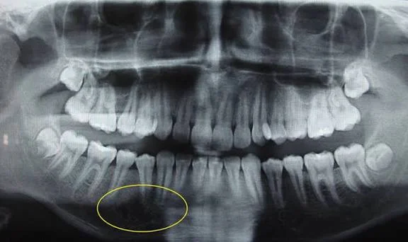
Jaw cysts are biologically insidious. During early intraosseous growth, they produce zero pain or visible swelling. However, as the lesion expands, prominent jawbone cyst symptoms emerge:
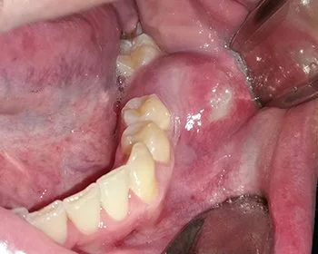
- Asymptomatic Expansion and Bony Bulging: Hard or springy swelling detectable upon palpation as the cyst thins the outer cortical bone plate.
- Tooth Displacement and Mobility: The expanding fluid sac pushes adjacent tooth roots, causing healthy teeth to tilt, drift, or become mobile.
- Discoloration and Loss of Tooth Vitality: Pressure exerted on neighboring root apices can sever neurovascular supply, causing teeth to lose vitality and darken.
- Lip and Chin Numbness (Paresthesia): Particularly in lower jawbone cyst cases, pressure exerted on the inferior alveolar nerve canal causes localized numbness or tingling in the lower lip and chin.
- Pathologic Jaw Fractures: Severe bone erosion can weaken the mandible so significantly that minor chewing forces trigger an unexpected fracture.
- Secondary Infection: If the cyst becomes infected, patients experience sudden acute pain, facial swelling, bad taste, and purulent discharge.
Is a Jaw Cyst Dangerous?
Upon receiving a diagnosis, patients frequently ask is a jaw cyst dangerous?
The medical answer is straightforward: The vast majority (90% to 95%) of jaw cysts are benign, meaning they do not metastasize like malignant cancers. However, benign does not mean harmless. If left untreated, a jaw cyst can:
- Cause massive bone loss leading to spontaneous jaw fractures.
- Destroy adjacent healthy teeth and root structures.
- Compress the inferior alveolar nerve, causing permanent lip numbness.
- In rare instances, undergo neoplastic transformation within its epithelial lining into aggressive tumors (such as ameloblastoma).
Therefore, even benign jaw cysts present clinical risks and require surgical intervention.
How to Shrink a Jaw Cyst?
When managing massive cysts occupying large portions of the jaw or enveloping vital nerve structures, immediate complete enucleation carries a risk of nerve damage or jaw fracture. To address how to shrink a jaw cyst, surgeons deploy a staged technique called marsupialization:
- Decompression (Marsupialization): A small surgical window is created into the cyst wall, evacuating the fluid. A small tube or stent is sutured in place to keep the window open.
- Internal Pressure Relief: Removing hydrostatic pressure stops bone resorption and prompts the surrounding bone to regenerate. The cyst cavity shrinks significantly over several months.
- Secondary Enucleation: Once the cyst has shrunk away from vital nerves, a secondary minor procedure easily removes the remaining small cyst lining in one piece.
Surgical Removal: Lower Jawbone Cyst Surgery
Clinical presentation of a lower jawbone cyst requires meticulous planning due to the dense cortical bone of the mandible and its proximity to the inferior alveolar nerve. The steps of lower jawbone cyst surgery proceed as follows:
- 3D Volumetric Mapping: A 3D CBCT scan maps the exact spatial relationship between the cyst, the nerve canal, and adjacent teeth.
- Anesthesia: The procedure is performed under deep local anesthesia, IV sedation, or general anesthesia, depending on lesion size.
- Enucleation (Cyst Removal): A mucoperiosteal flap is reflected, and the thin overlying bone is removed. The cyst capsule is carefully peeled away intact from the surrounding bone bed and nerve canal.
- Cavity Curettage and Pathology: The bony walls are smoothed to eliminate microscopic epithelial remnants. The excised cyst tissue is immediately sent for histopathological examination.
- Bone Grafting (Graft Application): If the resulting defect is large, a synthetic or xenograft bone graft (bone powder) covered by a protective collagen membrane is placed to accelerate bone healing. The flap is closed with sutures.
Post-Operative Recovery: Jaw Cyst Surgery Recovery
To ensure smooth healing during your jaw cyst surgery recovery, observe these post-operative rules:
- Cold Compress: Apply external ice packs over the cheek for 10-minute intervals during the first 24 to 48 hours to minimize facial swelling.
- Dietary Care: Stick to a soft, lukewarm diet for the first week. Avoid hot, spicy, acidic, or crunchy foods, and chew on the unoperated side.
- Oral Hygiene: Use prescribed chlorhexidine mouthwashes starting the day after surgery, brushing unoperated teeth normally while avoiding trauma to the suture line.
- Protect the Jawbone: Avoid contact sports or biting into hard foods for a few months following large cyst removal to protect the healing bone from structural stress.
Comparative Matrix: Enucleation vs. Marsupialization
Compare the two primary surgical approaches for jaw cysts below:
|
Technical Criteria |
Surgical Enucleation (Single Stage) |
Marsupialization (Staged Decompression) |
|
Surgical Approach |
Entire cyst capsule peeled out in one piece |
Surgical window created to drain fluid via stent |
|
Number of Procedures |
Single Stage |
Two-Stage (Decompression followed by removal) |
|
Inferior Alveolar Nerve Protection |
Carries risk if cyst contacts nerve directly |
Minimizes risk of permanent nerve paresthesia |
|
Pathologic Fracture Risk |
Present during removal of massive cysts |
Low, as bone regenerates during decompression |
|
Overall Healing Window |
1 to 2 Weeks (Suture removal phase) |
3 to 9 Months (Active shrinkage phase) |
|
Biopsy Timing |
Entire tissue sent immediately for pathology |
Tissue sample taken during window creation |
Frequently Asked Questions
Will I feel any pain during jaw cyst surgery?
No. High-potency local anesthetic blocks, sedation, or general anesthesia ensure that the jawbone, teeth, and surrounding soft tissues are completely numbed. You will feel only slight tactile pressure or instrument vibrations during the surgery; pain signals are biologically blocked.
Does a bone cavity left by a jaw cyst heal on its own?
Small bone cavities remaining after cyst removal heal spontaneously as the body converts a stable blood clot into new bone. However, if the cyst created a large defect or thinned the cortical jaw boundaries, placing a jaw cyst bone graft during surgery is necessary to support bone regeneration and prevent structural collapse.
Can a jaw cyst recur after surgical removal?
Most common jaw cysts (such as radicular cysts) do not recur once the capsule is completely enucleated. However, aggressive cyst types like the Odontogenic Keratocyst (OKK) can leave microscopic satellite cysts within the bone, presenting a higher recurrence risk. Patients treated for complex cysts should be monitored with annual panoramic X-rays for 5 years.
When are sutures removed after jaw cyst surgery?
If non-resorbable micro-sutures were placed, they are gently and painlessly removed by your oral surgeon 7 to 10 days after surgery.
About the Author