


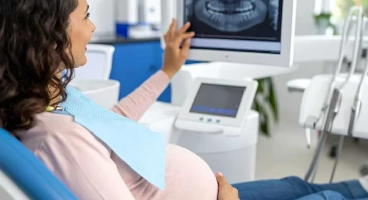
Dental X-Rays During Pregnancy: Safety, Fetal Risk and Shields
Don't put off your smile. Message us now to discuss your treatment options with our expert dentists.
WhatsAppGestational pregnancy initiates dramatic endocrine surges that heighten capillary permeability within the gingival architecture, alter salivary buffering capacity, and exacerbate acute marginal inflammation (pregnancy gingivitis). When expectant mothers experience acute odontogenic pain, deep pulpal decay, periapical abscesses, or impacted third molar infections, leaving these infectious sites untreated carries significant systemic risks; chronic bacteremia and inflammatory mediators circulating into systemic blood flows are clinically correlated with premature labor and low birth weight. However, no dental surgeon can ethically or safely perform invasive endodontic therapy or surgical debridement blind without definitive radiographic diagnostics.
The moment diagnostic radiation is mentioned, expectant parents frequently experience extreme anxiety. Persistent cultural myths suggesting that diagnostic dental rays cause fetal congenital malformations, teratogenesis, or spontaneous abortion induce understandable hesitancy. The primary clinical query raised by pregnant patients is whether dental X-rays during pregnancy are medically permissible. Patients routinely inquire: can you get dental X-rays during pregnancy, are dental X-rays during pregnancy harmful using digital sensors, is a panoramic dental X-ray during pregnancy safe, what to do if I had dental X-ray before knowing I was pregnant what should I do, how does a lead apron dental X-ray pregnancy protect the fetus, what are modern pregnancy dental radiographs guidelines, and fundamentally do dental X-rays harm the fetus? This clinical guide covers the radiobiological physics, dosimetric evaluations, and safety parameters governing obstetric dental imaging.
Table of Content
- Can You Get Dental X-Rays During Pregnancy?
- Are Dental X-Rays During Pregnancy Harmful?
- Protective Shielding: Lead Aprons and Thyroid Collars
- Panoramic Dental X-Ray During Pregnancy: Clinical Indications
- Accidental Exposure: Had Dental X-Ray Before Knowing I Was Pregnant What Should I Do?
- Radiographs Across Gestational Trimesters
- Do Dental X-Rays Harm the Fetus? Deconstructing Cultural Myths
- Comparative Matrix: Radiation Dosages in Medical Imaging & Nature
- Frequently Asked Questions
Can You Get Dental X-Rays During Pregnancy?
According to published clinical guidelines from the American Dental Association (ADA), the American College of Obstetricians and Gynecologists (ACOG), and the American College of Radiology (ACR): Yes, diagnostic dental radiographs are completely safe and medically authorized throughout all stages of pregnancy when protective shielding is utilized.
Why is this medical guidance clear and unequivocal?
- Collimated Craniofacial Trajectory: Dental X-ray tubes utilize focused cylindrical collimators aimed exclusively at the maxilla, mandible, or single dental crowns. The primary beam path is directed toward oral hard tissues, anatomically distant from the gravid uterus and developing fetus.
- Microscopic Scatter Exposure: The ionizing radiation dose required to expose modern digital sensors or ultra-speed E/F dental film is minuscule compared to general medical computed tomography (CT) scans of the abdomen or pelvis.
- The Clinical Risk of Untreated Sepsis: An acute, throbbing periapical abscess releasing bacterial endotoxins and inducing maternal systemic pyrexia presents a substantially greater biological hazard to the developing pregnancy than the minute exposure of a single localized radiograph.
Are Dental X-Rays During Pregnancy Harmful?
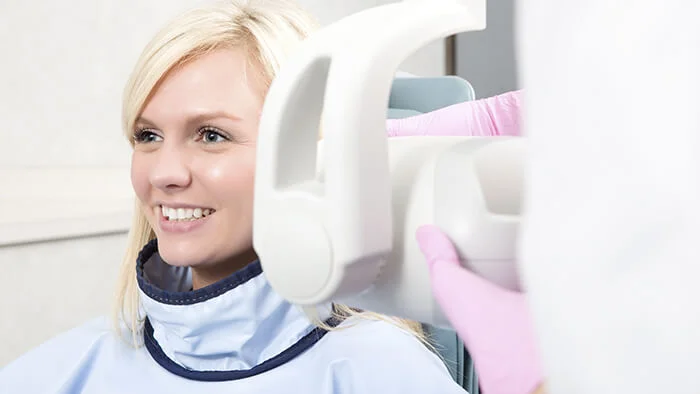
Evaluating whether are dental X-rays during pregnancy harmful requires reviewing radiation dosimetry measured in microsieverts:
- Established Teratogenic Threshold for the Fetus: The International Commission on Radiological Protection (ICRP) and ACOG confirm that fetal structural malformations, microcephaly, or growth restriction require a direct cumulative uterine exposure exceeding fifty thousand microsieverts (fifty milligray).
- Single Digital Periapical Radiograph: Emits approximately 2 to 5 microsieverts at the primary craniofacial target. The secondary scatter radiation reaching the shielded pelvic region is virtually immeasurable, typically falling below zero point one microsievert.
- Daily Natural Environmental Background Radiation: Every human on earth absorbs approximately eight to ten microsieverts of natural background radiation daily from cosmic rays, mineral deposits in soil, and ambient radon gas.
- Commercial Transatlantic Flight Comparison: An expectant mother flying a round trip across international flight paths absorbs forty to fifty microsieverts of high-altitude cosmic exposure, comparable to receiving ten to twenty localized dental periapical films.
Therefore, the biological likelihood of a single diagnostic digital dental radiograph causing fetal harm is negligible.
Protective Shielding: Lead Aprons and Thyroid Collars
In modern dental centers, adhering to the ALARA (As Low As Reasonably Achievable) radiation safety principle means utilizing full physical barrier shielding for every obstetric patient:
- Lead Apron Shielding: Draped across the patient from the clavicles down through the knees, the lead apron provides a high-density metallic barrier. It absorbs secondary scatter photons, preventing scatter radiation from reaching the pelvic cavity and gravid uterus.
- Thyroid Collar Shielding: Encircling the anterior cervical neck, the lead collar protects maternal thyroid follicles from scatter, preventing metabolic endocrine disruption.
- Collimated Digital Photostimulable Sensors: Modern digital CMOS and CCD sensors require up to eighty percent less radiation time compared to legacy film packets, further minimizing total photon exposure.
Panoramic Dental X-Ray During Pregnancy: Clinical Indications
Patients routinely ask whether a wide-angle panoramic dental X-ray during pregnancy can be taken:
- An extraoral panoramic radiograph rotates around the patient's cranium, capturing all maxillary and mandibular teeth, both temporomandibular joints, and the maxillary sinuses in a single sweeping scan.
- Total digital exposure measures approximately ten to fifteen microsieverts, still thousands of times below the fifty thousand microsievert fetal danger threshold.
- However, obstetric protocols emphasize targeted diagnostic discipline. Instead of broad extraoral scans, clinicians prioritize focused periapical intraoral films that capture only the problematic tooth.
- Unless required for acute maxillofacial trauma, severe deep facial cellulitis, or emergency surgical extractions, comprehensive panoramic surveys are typically deferred until after delivery.
Accidental Exposure: Had Dental X-Ray Before Knowing I Was Pregnant What Should I Do?
A frequent, highly emotional scenario presented to obstetricians is: had dental X-ray before knowing I was pregnant what should I do?
- Remain Calm and Reassured: Do not consider elective termination of a desired pregnancy because of one or two dental radiographs taken prior to a positive pregnancy test.
- The "All-or-None" Radiobiological Principle: During the pre-implantation and early organogenesis period (the first two to three weeks following conception), blastocyst cells are undifferentiated. Under the radiobiological "all-or-none" law, severe exposures (such as pelvic radiation therapy) disrupt implantation; conversely, if the pregnancy continues, undamaged blastomeres compensate fully, resulting in normal embryogenesis.
- The scatter exposure from a localized dental film does not approach embryotoxic levels. Inform your obstetrician during your initial prenatal examination, review the event, and proceed with standard ultrasound screenings with confidence.
Radiographs Across Gestational Trimesters
Managing diagnostic imaging across gestational trimesters follows established obstetric timing guidelines:
1. First Trimester (Weeks 1 through 12)
This critical organogenesis phase involves rapid embryonic tissue differentiation and organ development. Elective cosmetic smile makeovers, elective veneers, and routine checkups are deferred. However, if emergency toothache, pulpitis, or acute dentoalveolar abscesses emerge, localized diagnostic radiographs with lead shielding must proceed to diagnose the source and deliver emergency treatment.
2. Second Trimester (Weeks 13 through 27)
Universally recognized as the optimal, safest window for dental treatment. Maternal organogenesis is complete, nausea has resolved, and the physical size of the uterus permits comfortable positioning in the dental chair. Necessary operative restorations, root canal treatments, urgent surgical extractions, and indicated periapical films proceed smoothly.
3. Third Trimester (Weeks 28 to Delivery)
While fetal organs are fully formed, extended supine positioning in a dental chair can compress the maternal inferior vena cava, inducing supine hypotensive syndrome. Radiographs are reserved for acute dental emergencies requiring rapid relief before labor.
Do Dental X-Rays Harm the Fetus? Deconstructing Cultural Myths
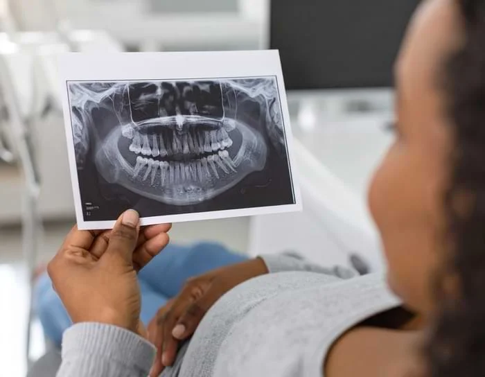
Addressing common misconceptions helps dispel unnecessary fear:
- Myth: "A routine tooth X-ray causes mental developmental delay or limb malformations."
Fact: Fetal teratogenicity requires ionizing radiation exposures exceeding fifty thousand microsieverts delivered directly to the pelvic region. A localized dental film generates negligible scatter, posing zero risk of physical malformation.
- Myth: "Enduring intense toothache is safer for the baby than having an X-ray taken."
Fact: Maternal systemic stress, elevated cortisol, untreated fever, and bacteremia from dental abscesses elevate risks of premature labor. Prompt radiographic diagnosis is the safer clinical choice.
- Myth: "Radiation passes into breast milk and poisons the nursing infant."
Fact: X-rays are electromagnetic waves traveling at the speed of light; they pass through matter and vanish instantly upon deactivating the beam. No radiation residues accumulate in maternal bodily fluids or breast milk.
Comparative Matrix: Radiation Dosages in Medical Imaging & Nature
|
Ionizing Radiation Source |
Typical Effective Dose |
Fetal Teratogenic Danger Rating |
|
Established Threshold for Fetal Teratogenic Harm |
50,000 Microsieverts (50 mGy) |
Hazard Threshold |
|
1 Single Digital Intraoral Periapical X-Ray |
2 to 5 Microsieverts |
Negligible (Zero Documented Risk) |
|
1 Single Digital Panoramic Dental X-Ray |
10 to 15 Microsieverts |
Inconsequential |
|
Daily Natural Ambient Background Radiation |
8 to 10 Microsieverts daily |
Baseline biological background |
|
Transatlantic Passenger Flight (London to New York) |
40 to 50 Microsieverts |
Standard commercial travel |
|
Standard Diagnostic Chest Radiograph (CXR) |
80 to 100 Microsieverts |
Clinically safe threshold |
Frequently Asked Questions
Is wearing a lead apron absolutely mandatory during pregnancy for a dental X-ray?
Yes. Although modern digital dental X-rays deliver minuscule scatter doses, international radiation safety protocols and clinical regulations make the application of a dual-sided lead apron and thyroid shield mandatory for any confirmed or suspected pregnancy.
Can an ultrasound or MRI be substituted for a dental X-ray during pregnancy?
No. Diagnostic ultrasound and magnetic resonance imaging (MRI) excel at detailing soft tissue structures, but they cannot resolve mineralized enamel crystalline structures, microscopic root canal anatomy, or early crestal bone loss. Ionizing X-rays remain the only diagnostic modality capable of imaging dental hard tissues accurately.
Is it safe to breastfeed immediately after receiving a dental X-ray?
Yes, completely safe. Diagnostic X-rays do not render human tissue, blood, or breast milk radioactive. Once the exposure button is released, photons dissipate entirely. Mothers can nurse immediately without pumping or discarding milk.
Can a dentist perform an endodontic root canal without taking an X-ray?
Simple restorative fillings can occasionally be prepared based solely on direct visual and tactile examination. However, performing endodontic therapy requires at least one initial periapical radiograph to accurately determine working length, evaluate root curvature, and confirm the absence of root fractures; proceeding without imaging risks procedural complications.
About the Author