


What Is a Salivary Gland Stone? Symptoms and Treatment Methods
Don't put off your smile. Message us now to discuss your treatment options with our expert dentists.
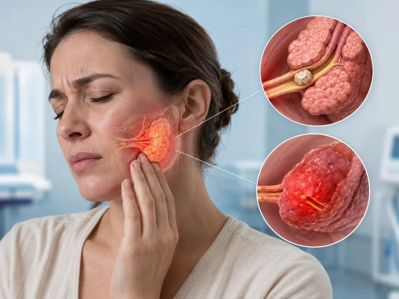
WhatsAppTo facilitate digestion, buffer cariogenic bacterial acids, lubricate the oral mucosa, and aid speech articulation, the human body secretes approximately one to one and a half liters of saliva each day. Saliva is produced by three paired major glands, the parotid in front of the ears, the submandibular beneath the lower jaw, and the sublingual under the floor of the mouth, alongside hundreds of microscopic minor glands. Clear saliva travels through narrow excretory ducts into the oral cavity. However, chronic systemic dehydration, reduced salivary flow velocity, or altered salivary chemistry can cause calcium phosphate and calcium carbonate salts to precipitate and form mineralized deposits. Clinically termed sialolithiasis, this calcification is universally known as a salivary gland stone.
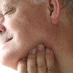
Experiencing acute jaw swelling that enlarges like an egg during meals accompanied by tight throbbing discomfort causes significant patient anxiety.
Table of Content
- What Is a Salivary Gland Stone?
- What Causes Salivary Gland Stone? Primary Etiological Drivers
- Salivary Gland Stone Symptoms: Recognizing Clinical Indicators
- Does Salivary Gland Stone Pass on Its Own?
- Salivary Gland Stone Passing Methods: Conservative Home Relief
- Salivary Gland Stone Treatment: Removal Techniques
- Comparative Matrix: Sialolithiasis Treatment Approaches
- Frequently Asked Questions
What Is a Salivary Gland Stone?
In maxillofacial surgery, a salivary gland stone is defined as a calcified intraductal or intraparenchymal structure formed by the incremental concentric crystallization of calcium salts around an initial organic nidus (such as desquamated epithelial cells, mucus plugs, or bacterial colonies).
Ranging from small granules to large calcifications over two centimeters, stones that obstruct salivary ducts prevent normal fluid escape. The resulting retrograde pressure stretches the fibrous capsule of the gland, triggering severe inflammatory edema and pain. Approximately eighty to eighty-five percent of all stones develop within the submandibular gland system (Wharton's duct). This predilection occurs because Wharton's duct runs an upward trajectory against gravity, spans a longer distance, and carries saliva containing higher concentrations of mucin, calcium, and alkaline pH.
What Causes Salivary Gland Stone? Primary Etiological Drivers
When evaluating what causes salivary gland stone, clinical studies highlight several primary contributing variables:
- Systemic Dehydration: Chronic inadequate oral fluid intake concentrates salivary electrolytes, reducing liquid volume and promoting calcium precipitation.
- Salivary Stasis (Reduced Flow Rate): Stagnant intraductal saliva allows mineral salts to settle onto organic matrices rather than flushing out.
- Anatomical Course and Gravity: The submandibular duct ascends toward the anterior floor of the mouth; this upward gradient slows fluid clearance, facilitating sedimentation.
- Anticholinergic and Diuretic Medications: Antihypertensives, systemic diuretics, antihistamines, and psychiatric medications reduce salivary gland production.
- Ductal Micro-Trauma and Strictures: Accidental biting or mechanical trauma to the ductal papilla can induce fibrotic narrowing that traps crystalline particles.
- Altered Mineral Metabolism: Elevated serum calcium levels or hyperuricemia associated with gout can occasionally predispose to calculus formation.
Salivary Gland Stone Symptoms: Recognizing Clinical Indicators

The most characteristic salivary gland stone symptoms are closely linked to gustatory stimulation:
- Postprandial Sudden Glandular Swelling: Anticipating food, tasting sour items like lemons, or chewing stimulates parasympathetic salivary secretion. Because the calcification blocks outflow, saliva accumulates behind the obstruction; within minutes, a firm swelling expands under the jaw or in front of the ear.
- Gradual Resolution Between Meals: One to two hours after eating, salivary secretion slows, small volumes leak around the obstruction, and swelling slowly subsides. This cyclical pattern is typical of sialolithiasis.
- Referred Pain Patterns: Intraductal distension radiates throbbing pain upward to the ear, temple, and lower mandibular border.
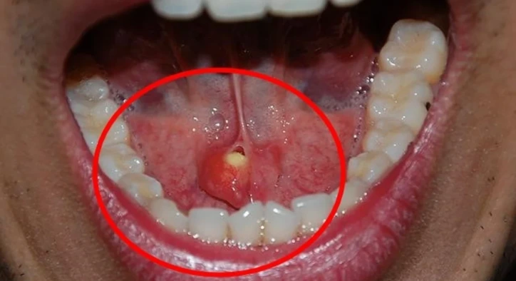
- Sublingual Salivary Gland Stone Presentation: A palpable, yellowish-white firm nodule detectable with the tongue along the floor of the mouth.
- Suppuration and Foul Taste (Sialadenitis): Secondary retrograde bacterial contamination can produce foul-tasting, purulent discharge into the mouth, accompanied by localized fever.
Does Salivary Gland Stone Pass on Its Own?
Addressing does salivary gland stone pass on its own depends primarily on stone dimensions and position:
- Spontaneous Clearance: Stones measuring two to three millimeters located close to the ductal orifice may occasionally flush out naturally under high salivary pressure aided by hydration and massage.
- Non-Passing Stones: Stones larger than four to five millimeters lodged in deep ductal bends or within the parenchymal body cannot negotiate narrower distal duct segments independently and require specialist intervention.
Salivary Gland Stone Passing Methods: Conservative Home Relief
For small stones, conservative salivary gland stone passing methods and approaches for how to get rid of salivary gland stone include:
- Aggressive Hydration: Consuming two and a half to three liters of plain water daily to maximize fluid output and hydrostatic pressure.
- Natural Sialogogue Stimulation: Sucking on fresh lemon wedges, sour candy, or chewing gum triggers salivary reflexes, creating hydraulic force behind the obstruction.
- Moist Heat and Directional Massage: Applying warm compresses over the submandibular region followed by gentle milking massage from back to front along the jawline toward the anterior floor of the mouth helps advance small stones.
Salivary Gland Stone Treatment: Removal Techniques
When conservative measures fail, structured salivary gland stone treatment is employed to address how salivary gland stone is removed:
1. Salivary Gland Stone Non Surgical Treatment (Interventional Sialendoscopy)
Sialendoscopy is the modern minimally invasive gold standard:
- Semi-rigid micro-endoscopes measuring less than one millimeter enter the duct orifice directly without external incisions.
- High-definition cameras locate the stone within the duct lumen.
- Miniature wire retrieval baskets or micro-forceps grasp and extract the stone under direct visualization.
- Larger stones are fragmented using holmium laser fibers or pneumatic lithotripters before extraction.
- The natural gland remains intact, no external scars are created, and patients return home the same day.
2. Transoral Papillotomy (Duct Slitting)
For stones impacted near the duct opening beneath the tongue, a minor incision under local anesthesia unroofs the duct, allowing extraction with forceps within minutes.
3. Salivary Gland Stone Surgery (Submandibular Sialoadenectomy)
When giant stones embed deep within the parenchyma, or chronic secondary infections cause permanent glandular fibrosis, open salivary gland stone surgery is indicated:
- Under general anesthesia, a small neck incision allows complete surgical excision of the compromised submandibular gland.
- Because the remaining major and minor salivary glands compensate fully, removing one submandibular gland does not induce chronic dry mouth.
Comparative Matrix: Sialolithiasis Treatment Approaches
|
Modality |
Stone Size & Location |
Incision / Anesthesia |
Recovery Timeline |
Gland Preservation |
|
Conservative Hydration |
Under 3 mm, distal duct |
None / None |
Immediate (1-3 days) |
100% preserved |
|
Interventional Sialendoscopy |
4 to 8 mm, accessible duct |
No incision / Local or sedation |
Same-day discharge |
100% preserved |
|
Transoral Papillotomy |
Proximal to sublingual papilla |
Minor intraoral incision / Local |
2 to 4 days |
100% preserved |
|
Open Gland Sialoadenectomy |
Large stones deep inside gland |
Cervical neck incision / General |
7 to 10 days |
Damaged gland removed |
Frequently Asked Questions
Can a salivary gland stone turn into cancer?
No. A salivary gland stone is a benign mineral calcification composed of calcium salts and protein matrix. It possesses no neoplastic or malignant potential. However, persistent untreated obstruction can lead to recurrent abscesses and chronic tissue scarring.
Is there a risk of facial nerve paralysis during salivary surgery?
The facial nerve branches control facial animation. The main facial nerve traverses the parotid gland, while the marginal mandibular branch runs near the submandibular gland. When performed by experienced maxillofacial or ENT surgeons utilizing continuous nerve monitoring, the risk of permanent nerve injury is low.
What happens if an obstructed salivary stone is left untreated?
Stagnant saliva behind an obstruction serves as a breeding ground for bacteria. Untreated sialolithiasis can escalate into deep neck space infections (Ludwig's angina), abscesses, and irreversible parenchymal tissue destruction.
Can lemon juice dissolve a salivary gland stone?
No. Ingesting lemon juice or vinegar cannot chemically dissolve calcium stones within the duct. The therapeutic value of lemon lies entirely in triggering a salivary reflex, generating hydrostatic fluid pressure to dislodge small stones.
About the Author