


What Is Dental Curettage? How Is It Performed, and Is It Painful?
Don't put off your smile. Message us now to discuss your treatment options with our expert dentists.
WhatsAppWhen periodontal inflammation is not halted at the reversible gingivitis stage, pathogenic subgingival biofilms and calculus deposits migrate apically along the root surface. This process destroys junctional epithelial attachments and periodontal ligament fibers, forming pathological anatomical spaces known as "periodontal pockets." Within pockets deeper than 4 millimeters, inaccessible to manual toothbrushes, interdental flossing, or supragingival scaling, anaerobic bacterial colonies release destructive endotoxins, driving progressive alveolar bone resorption.
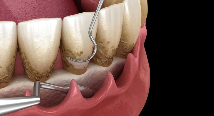
The primary non-surgical intervention for arresting deep periodontal breakdown and preventing tooth loss is dental curettage (subgingival scaling, root planing, and soft tissue debridement). Unlike conventional dental prophylaxis, this procedure meticulously debrides chronically inflamed pocket linings and removes micro-rough necrotic cementum from root surfaces.
Table of Content
- What Is Curettage and What Is Dental Curettage?
- Classifications of Subgingival Curettage
- How Dental Curettage Is Done?
- Dental Curettage Before and After: Tissue Responses
- Recovery After Dental Curettage and Pain Management
- What to Consider After Dental Curettage: Post-Op Care
- Comparative Matrix: Standard Scaling vs. Subgingival Curettage
- Frequently Asked Questions
What Is Curettage and What Is Dental Curettage?
In periodontal literature, the clinical definition of what is curettage is: The systematic removal of diseased subgingival granulation tissue, subgingival calculus, and endotoxin-saturated root cementum using specialized Gracey curettes, ultrasonic scalers, or dental lasers to facilitate soft tissue re-attachment and pocket reduction.
What clinical outcomes does a curettage treatment achieve?
- Infected Granulation Tissue Evacuation: Clears chronically inflamed sulcular pocket linings.
- Root Planing: Smooths microscopic root roughness, depriving bacterial biofilms of mechanical niches.
- Pocket Depth Reduction: Promotes shrinkage of 6 to 9 mm deep pockets down to healthy 2 to 3 mm maintenance levels.
- Arrests Alveolar Bone Loss: Eliminates persistent chronic inflammatory stimuli, stabilizing surrounding alveolar architecture.
- Reduces Tooth Mobility: Enables newly tightened gingival fibers to splint moderately mobile teeth naturally.
Classifications of Subgingival Curettage
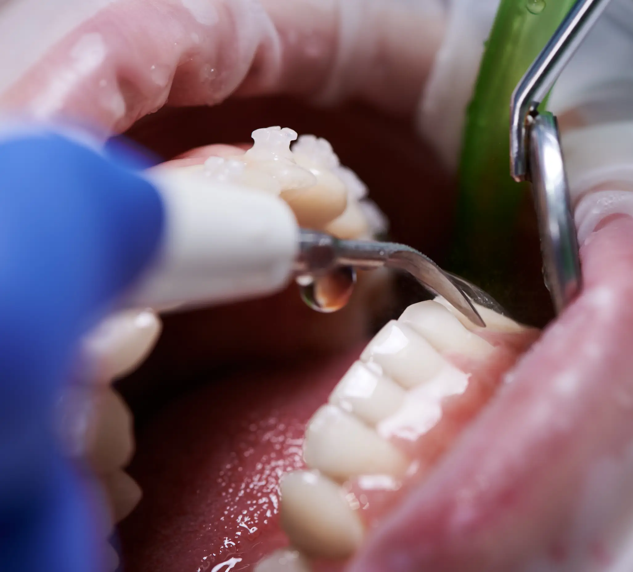
Depending on the anatomical severity of bone loss, curettage is categorized into two modalities:
1. Closed Subgingival Curettage (Non-Surgical Deep Scaling)
Indicated for moderate periodontitis with pocket depths measuring between 4 and 6 millimeters. Performed under local anesthesia without incising the gingiva, specialized curettes navigate beneath the gumline to debride root surfaces. The dentition is typically divided into quadrants and treated across 2 to 4 appointments.
2. Open Flap Curettage (Surgical Periodontal Therapy)
Indicated when pockets exceed 6 millimeters with complex infrabony defects. Under local anesthesia, the gingival tissue is surgically reflected (flap elevation) to provide direct visual access to deep root surfaces and jagged bone margins. Bone grafts and regenerative membranes may be placed before suturing.
How Dental Curettage Is Done?
Understanding how dental curettage is done involves five standard clinical phases:
- Periodontal Charting & Radiographic Analysis: Pocket depths are mapped across six sites per tooth using a calibrated periodontal probe, paired with digital radiographs.
- Local Anesthesia Administration: Local anesthetic is delivered to ensure the entire treatment area is completely numb and pain-free.
- Subgingival Debridement & Root Planing: Precision-angled Gracey curettes remove subgingival calculus and plane root surfaces smooth.
- Antiseptic Lavage & Laser Sterilization: The debrided pocket is irrigated with chlorhexidine or sterile saline, followed by diode laser decontamination where indicated.
- Tissue Compression & Hemostasis: Gentle digital compression adapts the gingival collar against the clean root surface, promoting stable blood clot formation.
Dental Curettage Before and After: Tissue Responses
Documented dental curettage before and after clinical transitions reveal significant physiological improvements:
- Pre-Treatment Status: Dark red or purplish gingiva, spontaneous bleeding upon probing, purulent discharge, chronic malodor, and deep pockets.
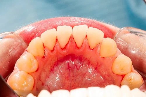
- Post-Treatment Status: Within 2 to 3 weeks, tissues transition into a firm, coral-pink architecture with zero bleeding on probing and tightened pocket margins.
- Apparent Root Exposure: As inflamed, swollen gingiva resolves and contracts, root surfaces may become slightly visible. This tissue recession is not a procedural defect; it reflects genuine resolution of inflammatory edema.
Recovery After Dental Curettage and Pain Management
Understanding recovery after dental curettage involves standard biological adaptation phases:
- First 24 to 48 Hours: Mild tenderness or a dull ache may occur as local anesthesia dissipates, which is easily managed with mild over-the-counter analgesics like acetaminophen or ibuprofen.
- Transient Thermal Sensitivity: Clean, newly exposed root cementum may react temporarily to cold fluids or air for 1 to 2 weeks. Desensitizing toothpastes accelerate comfort.
- Healing Timetable: Initial soft tissue epithelial closure occurs within 7 to 14 days, while complete maturation of the long junctional epithelium connection requires 6 to 8 weeks.
What to Consider After Dental Curettage: Post-Op Care
To safeguard clinical outcomes, adhere strictly to what to consider after dental curettage:
- Initial Eating Window: Avoid eating or chewing until local anesthesia has completely worn off to prevent tongue or lip laceration.
- First-Day Diet: Consume lukewarm, soft foods for the first 24 hours; strictly avoid boiling soups, spicy seasonings, or abrasive crusts.
- Tobacco Cessation: Avoid smoking for at least 48 hours. Tobacco toxins constrict microvasculature and impede tissue re-attachment.
- Gentle Oral Hygiene: Resume gentle brushing using an ultra-soft brush the following day, paired with prescribed antimicrobial chlorhexidine rinses.
- Daily Interdental Cleaning: Clean enlarged interdental spaces daily with appropriately sized interdental brushes once healing begins.
Comparative Matrix: Standard Scaling vs. Subgingival Curettage
|
Clinical Parameters |
Standard Dental Scaling (Prophylaxis) |
Subgingival Curettage (Deep Planing) |
|
Anatomical Domain |
Coronal to gingival margin (Supragingival) |
Deep within periodontal pockets (Subgingival) |
|
Anesthetic Requirement |
Rarely requires local anesthesia |
Mandatory local anesthesia (100% painless) |
|
Instrumentation |
Ultrasonic scaler tips |
Specialized Gracey curettes, lasers, hand tools |
|
Target Tissue |
Calcified calculus and external stains |
Subgingival calculus and granulation tissue |
|
Primary Goal |
Gingivitis prevention and stain removal |
Arresting alveolar bone loss and saving teeth |
|
Appointment Structure |
Single session (30-45 minutes) |
Usually 2 to 4 quadrant-based sessions |
Frequently Asked Questions
Does dental curettage hurt?
No. Local anesthesia is administered before the procedure begins, completely numbing the treatment area. Patients feel only mild physical pressure and instrument movements during treatment, without sharp pain.
What happens if recommended curettage is avoided?
Avoiding necessary curettage allows subgingival anaerobic bacteria to continue destroying the alveolar bone. Over time, periodontal pockets deepen, teeth become loose, recurrent periodontal abscesses develop, and non-decayed natural teeth will eventually require extraction.
Does curettage cause gaps between teeth?
Curettage does not create gaps. However, as chronically swollen, inflamed gums heal, they shrink down to their natural healthy dimensions. This shrinkage exposes pre-existing spaces where bone and tissue had already been lost to disease, signifying successful healing.
How often does dental curettage need to be repeated?
With diligent home hygiene and routine 6-month supportive periodontal cleanings, curettage often does not need to be repeated. If oral hygiene is neglected, pockets may reinfect, necessitating re-treatment after several years.
About the Author